 Ovarian Tumors What is the clinical setting when you will consider an ovarian mass? You would consider an ovarian mass, in any woman who comes in complaining of pressure symptoms. These symptoms include urinary frequency, pelvic discomfort, and ...
What patients and caregivers need to know about. If you or someone you love has cancer, know what to expect can help you cope. From basic information about cancer and its causes to detailed information about specific types of cancer, including risk factors, early detection, diagnosis and treatment options, you will find it here. More Topics You can help reduce the risk of cancer by making healthy choices like eating well, staying active and not smoking. It is also important to follow recommended screening guidelines, which can help detect certain early cancers. More Topics Cancer Prevention Tools Whether you want to learn about treatment options, get tips on how to deal with side effects, or have questions about health insurance, we are here to help. We can even find a free trip to treatment or a free place to stay when the treatment is away from home. More Top History NAVIGATION WORKING Top Our team of expert journalists brings you all the angles of cancer history – from last-minute news and survivor stories to detailed information about cutting-edge research. More news about Top Story cancer What is needed to overcome cancer? Research. We've invested more than $5 billion in cancer research since 1946, all to find more treatments, discover factors that can cause cancer and improve the quality of life of cancer patients. Life Saving Research Explore Our ResearchWe couldn't do what we do without our volunteers and donors. Together, we are making a difference – and you can too. Become a volunteer, make a deductible tax donation, or participate in a fundraising event to help us save lives. MORE MEASURES TO INVOLVATEOur PassionThe partners highlight the Health Care of the NFLPamperado Chef PartnershipJoin GrowNation The American Cancer Society was unable to do what we do without the support of our partners. Learn more about these partnerships and how you can also join us in our mission to save lives, celebrate lives and lead the struggle for a cancer-free world. Cancer PartnersPartners SpotlightThe NFL Defender Health QuizPampered Chef PartnershipJoin GrowNation At the American Cancer Society, we are on a mission to free the world from cancer. Until we do, we will be funding and conducting research, sharing expert information, supporting patients and spreading the word on prevention. So you can live longer and better. Learn how to use a difference Tests for ovarian cancer If your doctor finds something suspicious during a pelvic exam, or if you have symptoms that may be caused by ovarian cancer, your doctor will recommend tests and tests to find the cause. Medical history and physical exam Your doctor will ask you about your medical history to know possible risk factors, including your family history. You will also be asked if you are having symptoms, when they started, and how long you have had them. Your doctor will likely do a pelvic exam to check if there is an enlarged ovary or signs of fluid in the abdomen (called ascites). If there are reasons to suspect that you have ovarian cancer based on your symptoms and/or physical exam, your doctor will order some tests to check later. Check with a specialist If your pelvic exam or other tests suggest you have ovarian cancer, you will need a doctor or surgeon who specializes in treating women with this type of cancer. A gynecological oncologist is an obstetrician/gynaecologist who is specially trained to treat cancers of the female reproductive system. Treatment by a gynecological oncologist helps ensure that you receive the best type of surgery for your cancer. It has also been shown to help patients with ovarian cancer live longer. Any person suspected of having ovarian cancer should see this type of specialist before being operated. Images Tests Doctors use imaging tests to take pictures of the inside of their body. The imaging tests may show whether a pelvic mass is present, but they cannot confirm that the mass is a cancer. These tests are also useful if your doctor is looking to see if ovarian cancer has spread (metasified) to other tissues and organs. Ultrasound (ultrasonography) uses sound waves to create an image on a video screen. The sound waves are released from a small probe placed in the woman's vagina and a small microphone-like instrument called a sound wave transducer and collects the echoes while bounced organs. A computer turns these echoes into an image on the screen. Ultrasound is often the first test performed if a problem with ovaries is suspected. It can be used to find an ovarian tumor and to check whether it is a solid mass (tumor) or a cyst full of fluid. It can also be used to get a better look at the ovary to see how big it is and how it looks inside. This helps the doctor decide which masses or cysts are more worrying. Computed tomography scans (CT) The X-ray test is a test that makes detailed cross section images of your body. The test can help determine whether ovarian cancer has spread to other organs. CT scans don't show small ovarian tumors well, but they can see larger tumors, and they can be able to see if the tumor is growing in nearby structures. Computed tomography can also find enlarged lymph nodes, signs of cancer spread to the liver or other organs, or signs that an ovarian tumor is affecting your kidneys or bladder. Tomographs are not usually used for biopsy of an ovarian tumor (see biopsy in the "Other Tests") section, but may be used for biopsy of a suspicious metastasis (dissemination area). For this procedure, called TC-guided needle biopsy, the patient remains on the CT scan table, while a radiologist moves a biopsy needle to the mass. CT scans are repeated until doctors trust that the needle is in the dough. A thin sample of needle biopsy (weather fragment) or a sample of nucleus needle biopsy (a thin 1⁄2 inch long tissue cylinder and less than 1/8 inches in diameter) is extracted and examined in the laboratory. Barium enema X-ray A barium enema is a test to see if the cancer has invaded the colon (big instino) or rectum. This test is rarely used for women with ovarian cancer. can be done instead. Magnetic resonance imaging (MRI) also creates cross section images of its interiors. But MRI uses strong magnets to make the images – not X-rays. A contrast material called gadolinium can be injected into a vein before exploration to see the details better. MRI scans are often not used to look for ovarian cancer, but they are particularly useful to examine the brain and spinal cord where cancer may spread. Torax X-ray An analysis can be done to determine whether ovarian cancer has spread (metasified) to the lungs. This spread can cause one or more tumors in the lungs and, more often, causes the fluid to recover around the lungs. This fluid, called pleural effusion, can be seen with chest x-rays and other types of scans. Postitron emission tomography (PET)For a radioactive glucose (sugar) is given to look for cancer. The body cells take different amounts of sugar, depending on how fast they are growing. Cancer cells, which grow rapidly, are more likely to absorb greater amounts of sugar than normal cells. A special camera is used to create an image of radiactivity areas in the body. The image of a PET scanner is not as detailed as a computed tomography or magnetic resonance, but provides useful information about whether abnormal areas seen in these other tests are likely to be cancer or not. If you have already been diagnosed with cancer, your doctor may use this test to see if the cancer has spread to the lymph nodes or other parts of the body. A PET analysis may also be useful if your doctor thinks that the cancer may have spread but does not know where. PET/CT scan: Some machines can make both a PET and a CT scan at the same time. This allows the doctor to compare areas of greater radioactivity in the PET analysis with the most detailed image of that area in the CT analysis. PET scans can help find cancer when it has spread, but are not often used to look for ovarian cancer. Other Laparoscopic Tests This procedure uses a thin, light tube through which a doctor can observe ovaries and other pelvic organs and tissues in the area. The tube is inserted through a small incision (cut) in the lower abdomen and sends the images of the pelvis or abdomen to a video monitor. Laparoscopy provides a vision of organs that can help plan surgery or other treatments and can help doctors confirm (how far the tumor has spread) of the cancer. In addition, doctors can manipulate small instruments through laparoscopic incision to perform biopsies. Colonoscopy A is a way to examine the inside of the large intestine (colon). The doctor looks at the entire length of the colon and rectum with a colonoscope, a thin, flexible and illuminated tube with a small video camera at the end. It is inserted through the anus and in the rectum and colon. Any abnormal area that is veda can be biopsy. This procedure is most commonly used to look for colorectal cancer. Biopsy The only way to determine for sure if a growth is cancer is to remove a piece of it and examine it in the laboratory. This procedure is called a . For ovarian cancer, biopsy is done more commonly by removing the tumor during surgery. In rare cases, a suspected ovary cancer may be biopsied during a laparoscopic procedure or a needle placed directly in the tumor through the skin of the abdomen. The needle will usually be guided by ultrasound or CT scan. This is done only if surgery is not performed due to advanced cancer or other serious medical conditions, as there is concern that a biopsy could spread cancer. If you have ascites (fluid construction within the abdomen), fluid samples may also be used to diagnose cancer. In this procedure, called paracentesis, the skin of the abdomen is numb and a needle attached to a syringe is passed through the abdominal wall to the fluid in the abdominal cavity. Ultrasound can be used to guide the needle. The fluid is absorbed in the syringe and then sent for analysis to see if it contains cancer cells. In all these procedures, the tissue or fluid obtained is sent to the laboratory. There it is examined by a pathologist, a doctor specializing in the diagnosis and classification of diseases by examining cells under a microscope and using other laboratory tests. Blood Tests Your doctor will order blood count tests to make sure you have enough red blood cells, white blood cells and platelets (cells that help stop the bleeding). There will also be tests to measure your kidney and liver function, as well as your overall health. The doctor will also order a CA-125 test. Women who have a high level of CA-125 often refer to a gynecological oncologist, but any woman with suspected ovarian cancer should see a gynecological oncologist, too. Some germ cell cancers can cause high blood levels of tumor markers of human choral gonadotropin (HCG), alpha-fetoprotein (AFP), and/or lactated dehydrogenase (LDH). These may be checked if your doctor suspects that your ovarian tumor may be a germ cell tumor. Some ovarian estromal tumors cause the blood levels of a substance called inhibition and hormones such as estrogen and testosterone to rise. These levels can be checked if your doctor suspects you have this type of tumor. Genetic advice and testing if you have ovarian cancer If you have been diagnosed with epithelial ovarian cancer, your doctor will likely recommend that you receive genetic advice and genetic tests for certain, even if you do not have a family history of cancer. The most common mutations are found in the BRCA1 and BRCA2 genes, but some ovarian cancers are in other genes such as ATM, BRIP1, RAD51C/RAD51D, MSH2, MLH1, MSH6, or PMS6. Genetic testing to search for inherited mutations may be useful in several ways You may have heard of some genetic testing at home. There is concern that these tests will be promoted by companies without giving full information. For example, a test for a small number has been approved by the FDA. However, there are more than 1,000 known BRCA mutations, and those included in the approved test are not the most common. This means that there are many BRCA mutations that would not be detected by this test. A genetic counselor or other qualified medical practitioner can help you understand the risks, benefits, and possible limits of what genetic tests can tell you. This can help you decide whether the test is right for you, and what tests are best. For more information on genetic testing, see Molecular Tests for Gene Changes In some cases of ovarian cancer, doctors may look for specific genetic changes in cancer cells (not blood or saliva samples) that might mean that certain may help treat cancer. These molecular tests can be done in a piece of cancer taken during a biopsy or surgery for ovarian cancer. BRCA1 and BRCA2 genetic mutations: BRCA genes are usually involved in DNA repair and mutations in these genes can keep DNA broken and cells cannot work properly. This can cause cells to grow out of control and become cancer. Some Ovarian cancers have BRCA gene mutations. MSI and MMR Gene Tests: Women who have clear cell, endometrioid or munic ovarian cancer may test their tumor to see if it shows high levels of genetic changes called microsatellite instability (MSI). Tests can also be done to see if cancer cells have changes in any of the desajustment repair genes (MMR) (MLH1, MSH2, MSH6, and PMS2). Changes in MSI or MMR genes (or both) are often seen in people with (HNPCC). Up to 10% of all ovarian epithelial cancers have changes in these genes. There are 2 possible reasons to test ovarian cancers for MSI or for MMR gene changes: NTRK gene mutations: Some ovarian cancers may be tested for changes in one of the NTRK genes. Cells with these genetic changes can lead to abnormal cell growth and cancer. Larotrectinib (Vitrakvi) and entrectinib (Rozlytrek) are targeted drugs that stop proteins made by NTRK abnormal genes. The number of ovarian cancers that have this mutation is very small, but this may be an option for some women. Our team consists of doctors and nurses certified oncology with deep knowledge of cancer care, as well as journalists, editors and translators with extensive medical writing experience. Chen, L., " Berek, J. (2018, January). UpToDate - Ovarian epithelial carcinoma, fallopian tube and peritoneum: clinical characteristics and diagnosis. Consultation on 6 February 2018, from https://www.uptodate.com/contents/epithelial-carcinoma-of-the-ovaryfallopian-tube-and-peritoneum-clinical-features-anddiagnosis.Coleman RL, Liu J, Matsuo K, Thaker PH, Weston SN and Sood Ak. Chapter 86: Carcinoma of the Fallopian Ovaries and Tubes. In: Niederhuber JE, Armitage JO, Doroshow JH, Kastan MB, Tepper JE, eds. Abeloff Clinical Oncology. Philadelphia, Pa. Elsevier: 2020.Konstantinopoulos PA, Norquist B, Lacchetti C, Armstrong D, Grisham RN, Goodfellow PJ, et al. Testing of twin and somatic tumors in epithelial ovarian cancer: ASCO Guideline. J Clin Oncol. 2020. doi: 10.1200/JCO.19.02960. [Epub in front of printing]. Koczkowska M, Zuk M, Gorczynski A, et al. Detection of somatic BRCA1/2 mutations in ovarian cancer - next generation sequencing analysis of 100 cases. Cancer Med. 2016;5(7):1640-1646. Morgan M, Boyd J, Drapkin R, Seiden MV. Ch 89 – Cancers that accumulate in the Ovary. In: Abeloff MD, Armitage JO, Lichter AS, Niederhuber JE, Kastan MB, McKenna WG,eds. Clinical oncology. 5th edition. Philadelphia, PA: Elsevier; 2014: 1592.National Integral Cancer Network (NCCN)—Genetic/Familial High Risk Assessment: Breast, Ovarian and Pancreatic. V1.2020. Access March 26, 2020 from https://www.nccn.org/professionals/physician_gls/pdf/genetics_bop.pdf Comprehensive Cancer Network (NNCN) - Ovarian Cancer including Autumn Tube and Primary Peritoneal Cancer. (2018, February 2). On February 5, 2018, from https://www.nccn.org/professionals/physician_gls/pdf/ovarian.pdf Comprehensive Cancer Network (NCCN) - Ovarian Cancer including Otope Tube Cancer and Primary Peritoneal Cancer. V1.2020. Access March 26, 2020 from http://www.nccn.org/professionals/physician_ gls/pdf/ovarian pdfOkamura R, Boichard A, Kato S, Sicklick JK, Bazhenova L, Kurzrock R. Analysis of NTRK Alterations in Malignities Pan-Cancer Adult and Pediatrics: Implications for NTR JCO Precis Oncol. 2018;2018:10.1200/PO.18.00183. doi:10.1200/PO.18.00183 Tewari KS, Penson RT and Monk BJ. Chapter 77: Ovarian Cancer. In: DeVita VT, Lawrence TS, Rosenberg SA, eds. DeVita, Hellman and Rosenberg Cancer: Principles " Oncology Practice. 11th edition. Philadelphia, Pa: Lippincott Williams " Wilkins; 2019.Weber S, McCann CK, Boruta DM, Schorge JO, Growdon WB. Laparoscopic Surgical staging of premature ovarian cancer. Reviews in Obstetrics and Gynaecology. 2011;4(3-4):117-122.Chen, L., & Berek, J. (2018, January). UpToDate - Ovarian epithelial carcinoma, fallopian tube and peritoneum: clinical characteristics and diagnosis. Consultation on 6 February 2018, from https://www.uptodate.com/contents/epithelial-carcinoma-of-the-ovaryfallopian-tube-and-peritoneum-clinical-features-anddiagnosis.Coleman RL, Liu J, Matsuo K, Thaker PH, Weston SN and Sood Ak. Chapter 86: Carcinoma of the Fallopian Ovaries and Tubes. In: Niederhuber JE, Armitage JO, Doroshow JH, Kastan MB, Tepper JE, eds. Abeloff Clinical Oncology. Philadelphia, Pa. Elsevier: 2020.Konstantinopoulos PA, Norquist B, Lacchetti C, Armstrong D, Grisham RN, Goodfellow PJ, et al. Testing of twin and somatic tumors in epithelial ovarian cancer: ASCO Guideline. J Clin Oncol. 2020. doi: 10.1200/JCO.19.02960. [Epub in front of printing]. Koczkowska M, Zuk M, Gorczynski A, et al. Detection of somatic BRCA1/2 mutations in ovarian cancer - next generation sequencing analysis of 100 cases. Cancer Med. 2016;5(7):1640-1646. Morgan M, Boyd J, Drapkin R, Seiden MV. Ch 89 – Cancers that accumulate in the Ovary. In: Abeloff MD, Armitage JO, Lichter AS, Niederhuber JE, Kastan MB, McKenna WG,eds. Clinical oncology. 5th edition. Philadelphia, PA: Elsevier; 2014: 1592.National Integral Cancer Network (NCCN)—Genetic/Familial High Risk Assessment: Breast, Ovarian and Pancreatic. V1.2020. Access March 26, 2020 from https://www.nccn.org/professionals/physician_gls/pdf/genetics_bop.pdf Comprehensive Cancer Network (NNCN) - Ovarian Cancer including Autumn Tube and Primary Peritoneal Cancer. (2018, February 2). On February 5, 2018, from https://www.nccn.org/professionals/physician_gls/pdf/ovarian.pdf Comprehensive Cancer Network (NCCN) - Ovarian Cancer including Otope Tube Cancer and Primary Peritoneal Cancer. V1.2020. Access March 26, 2020 from http://www.nccn.org/professionals/physician_ gls/pdf/ovarian pdfOkamura R, Boichard A, Kato S, Sicklick JK, Bazhenova L, Kurzrock R. Analysis of NTRK Alterations in Malignities Pan-Cancer Adult and Pediatrics: Implications for NTR JCO Precis Oncol. 2018;2018:10.1200/PO.18.00183. doi:10.1200/PO.18.00183 Tewari KS, Penson RT and Monk BJ. Chapter 77: Ovarian Cancer. In: DeVita VT, Lawrence TS, Rosenberg SA, eds. DeVita, Hellman and Rosenberg Cancer: Principles " Oncology Practice. 11th edition. Philadelphia, Pa: Lippincott Williams " Wilkins; 2019.Weber S, McCann CK, Boruta DM, Schorge JO, Growdon WB. Laparoscopic Surgical staging of premature ovarian cancer. Reviews in Obstetrics and Gynaecology. 2011;4(3-4):117-122. Last revision: April 3, 2020 American Cancer Society medical information is copyright material. For reprint requests, please see our . More In ovarian cancer Imagine a world free of cancer. Helps make it a reality. Cancer Information Programs & ServicesAbout ACSMore ACS SitesFollow us Cancer, Response, and Hope Information. Available every minute of every day. Follow Us© 2021 American Cancer Society, Inc. All rights reserved. The American Cancer Society is an organization exempted from qualified tax 501(c)(3). Tax identification number: 13-1788491. Cancer.org is a courtesy of the Leo family and Gloria Rosen. Select a Hope Lodge
Slideshow: Visual guide for ovarian cancer What is ovarian cancer? The current research suggests that this cancer begins in the fallopian tubes and moves to the ovaries, the twin organs that produce the eggs of a woman and the main source of female hormones estrogen and progesterone. Ovarian cancer treatments have become more effective in recent years, with the best results seen when the disease is early. Symptoms of Ovarian Cancer Early ovarian cancer rarely has symptoms. As the disease progresses, some symptoms may appear. These include: These symptoms may be caused by many conditions that are not cancer. If they occur persistently for more than a few weeks, report to your health care professional. Risk Factor: Family History A woman's chance of developing ovarian cancer is greater if a close relative has had ovarian, chest, or colon cancer. Researchers believe inherited genetic changes represent 10% of ovarian cancers. This includes mutations of the BRCA1 and BRCA2, which are linked to breast cancer. Women with a strong family history should talk to a doctor to see if closer medical follow-up might be helpful. Risk Factor: AgeThe strongest risk factor for ovarian cancer is age. It is very likely to develop after a woman passes through menopause. The use of postmenopausal hormone therapy can increase the risk. The link seems stronger in women who take estrogen without progesterone for at least 5 to 10 years. Doctors are not sure if taking a combination of estrogen and progesterone increases the risk as well. Risk Factor: Obesity Obese women have a higher risk of ovarian cancer than other women. And ovarian cancer mortality rates are also higher for obese women, compared to non-obese women. The heaviest women seem to have the greatest risk. Ovarian Cancer Screening There is no easy or reliable way to test ovarian cancer if a woman has no symptoms. No life-saving test has been shown when used in average-risk women. If you have symptoms that may be ovarian cancer, your doctor may suggest a rectavaginal pelvic exam, transvaginal ultrasound, or a CA-125 blood test to help find the cause. Diagnosis of ovarian cancerImage tests, such as ultrasounds or CT scan (see here), can help reveal an ovarian mass. But these scans cannot determine whether the anomaly is cancer. If cancer is suspected, the next step is usually surgery to remove suspicious tissues. A sample is then sent to the laboratory for further examination. This is called a biopsy. Ovarian Cancer Stages The initial surgery for ovarian cancer also helps to determine how far the cancer has spread, described by the following stages: Stage I: Confined to one or both ovaries Stage II: Divulgation to the uterus or other nearby organs Stage III: Disemination to the lymph nodes or abdominal liningStage IV: Disemination to distant liver organs These are malignant tumors that form from cells on the surface of the ovary. Some epithelial tumors are not clearly cancerous. These are known as tumors of low malignant potential (LMP). LMP tumors grow more slowly and are less dangerous than other forms of ovarian cancer. Ovarian Cancer Survival RatesOvarian cancer may be a terrifying diagnosis, with relative five-year survival rates ranging from 93% to 19% for epithelial ovarian cancer, depending on the stage of cancer. For LMP tumors, five-year relative survival rates range from 97 per cent to 89 per cent. Ovarian Cancer Surgery Surgery is used to diagnose ovarian cancer and determine its stage, but it is also the first stage of treatment. The goal is to eliminate as much of the cancer as possible. This may include a single ovary and nearby tissue at stage I. In more advanced stages, it may be necessary to remove both ovaries, along with the uterus and surrounding tissues. Chemotherapy In all stages of ovarian cancer, chemotherapy is usually given after surgery. This stage of treatment uses drugs to attack and kill any cancer that still remains in the body. Medicines may be given by mouth, through an intravenous pathway or directly in the abdomen (intraperitoneal chemotherapy). Women with LMP tumors generally do not need chemotherapy unless tumors grow after surgery. Targeted Therapies Researchers are working on therapies that aim at how ovarian cancer grows. A process called angiogenesis involves the formation of new blood vessels to feed tumors. A medicine called Avastin blocks this process, causing tumors to shrink or stop growing (see here in the illustration). After Treatment: Premature Menopause When women have both ovaries removed, they can no longer produce their own estrogen. This triggers menopause, no matter how young the patient is. The drop in hormone levels can also increase the risk of certain medical conditions, including osteoporosis. It is vital for women to be careful regularly after being treated for ovarian cancer. After Treatment: Moving OnWomen may find it takes a long time for your energy to come back after treatments are over. Fatigue is a very common problem after treatment for cancer. Starting a mild exercise program is one of the most effective ways to restore energy and improve emotional well-being. Check with your health care team to determine what activities are appropriate for you. Risk Reduction: PregnancyWomen who have biological children are less likely to have ovarian cancer than women who have never given birth. The risk seems to decrease with each pregnancy, and breastfeeding can provide additional protection. Risk reduction: 'Pildora Cancer'Ovaries is also less common in women who have taken birth control pills. Women who have used the pill for at least five years have about half the risk of women who never took the pill. Like pregnancy, birth control pills prevent ovulation. Some researchers think that ovulation can less often protect against ovarian cancer. Risk Reduction: Tubal ligationGetting tied tubes, officially known as tubal ligation, can offer some protection against ovarian cancer. The same thing happens to have a hysterectomy... removing the uterus. Risk Reduction: Removing ovaries For women with genetic mutations that put them at high risk for ovarian cancer, eliminating ovaries is an option. This can also be considered in women over 40 with a hysterectomy. Risk reducer: Low fat dietWhile there is no definitive diet to prevent ovarian cancer, there is evidence that what you eat can make a difference. In a recent study, women who get stuck on a low-fat diet for at least four years are less likely to develop ovarian cancer. Some researchers report that cancer is also less common in women who eat many vegetables, but more studies are needed. NextNext Slideshow Title1 Reviewed by IMAGES PROVIDED BY:1) 3D4Medical.com 2) Jupiterimages 3) Tom Merton/OJO Images 4) Eric Audras/PhotoAlto 5) Dirk Enters/Imagebroker.net 6) Adam Gault/OJO Images 7) Du Cane Medical Imaging Ltd. / Photo Researchers, Inc. 8) Kevin A. Somerville/Phototake 9) Professors Pietro M. Motta & Sayoko Makabe / Photo Researchers, Inc 10) Digital vision 11) ERProductions 12) Don Farrall/White 13) Photo taken 14) Digital vision 15) Tom Merton/OJO Images 16) Election of Christian Baitg/Photographer 17) Ian Hooton/Science Photo Library 18) Brain Evans/Photo Researchers Inc 19) LWA-Stephen Welstead/Flirt 20) Joff Lee/Fresh Food Images: American Cancer Society Website. Website of the National Cancer Institute. Website of the American Society of Menopause. This tool does not provide medical advice. This work does not prohibit medical help. It is intended for general information purposes only and does not address individual circumstances. It is not a substitute for professional medical advice, diagnosis or treatment and should not be based on making decisions about your health. Never ignore the professional medical advice in the search for treatment due to something you have read on the WebMD site. If you think you may have a medical emergency, call your doctor immediately or call 911. WebMD Slideshows See our presentations to learn more about your health. Top PicksHealth SolutionsMore WebMD Policies About WebMD Network Our applications for advertisers © 2005 - 2021 WebMD LLC. All rights reserved. DMA does not provide medical advice, diagnosis or treatment.

What 3D Ultrasound Can Tell You About Ovarian Cancer | Empowered Women's Health
Ovarian Cancer and Pregnancy | IntechOpen
Ovarian Tumors What is the clinical setting when you will consider an ovarian mass? You would consider an ovarian mass, in any woman who comes in complaining of pressure symptoms. These symptoms include urinary frequency, pelvic discomfort, and ...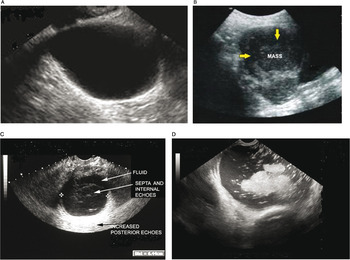
Ultrasound imaging of ovarian cancer (Chapter 22) - Ultrasonography in Gynecology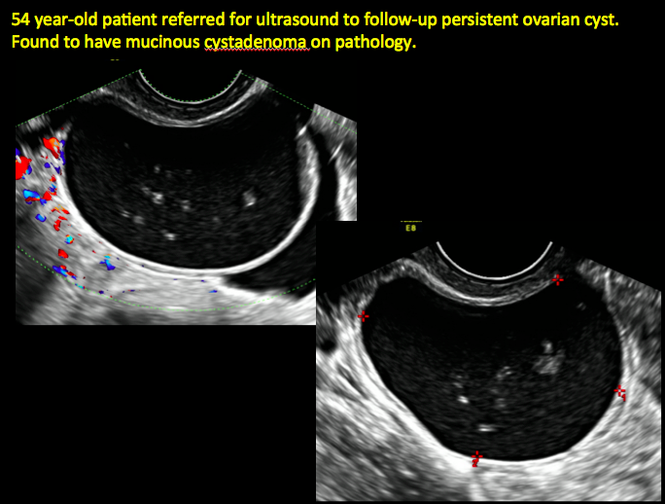
Imaging the suspected ovarian malignancy: 14 cases | MDedge ObGyn
Imaging Strategy for Early Ovarian Cancer: Characterization of Adnexal Masses with Conventional and Advanced Imaging Techniques | RadioGraphics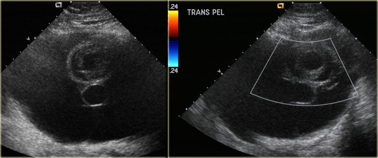
The Radiology Assistant : Common ovarian cystic lesions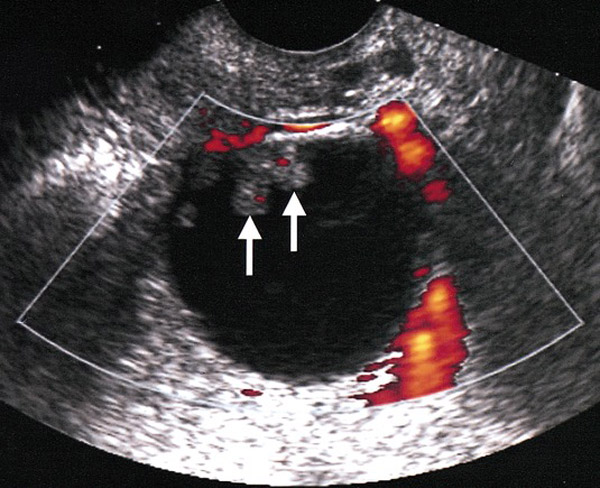
Ovarian Cancer - Oncology - Medbullets Step 2/3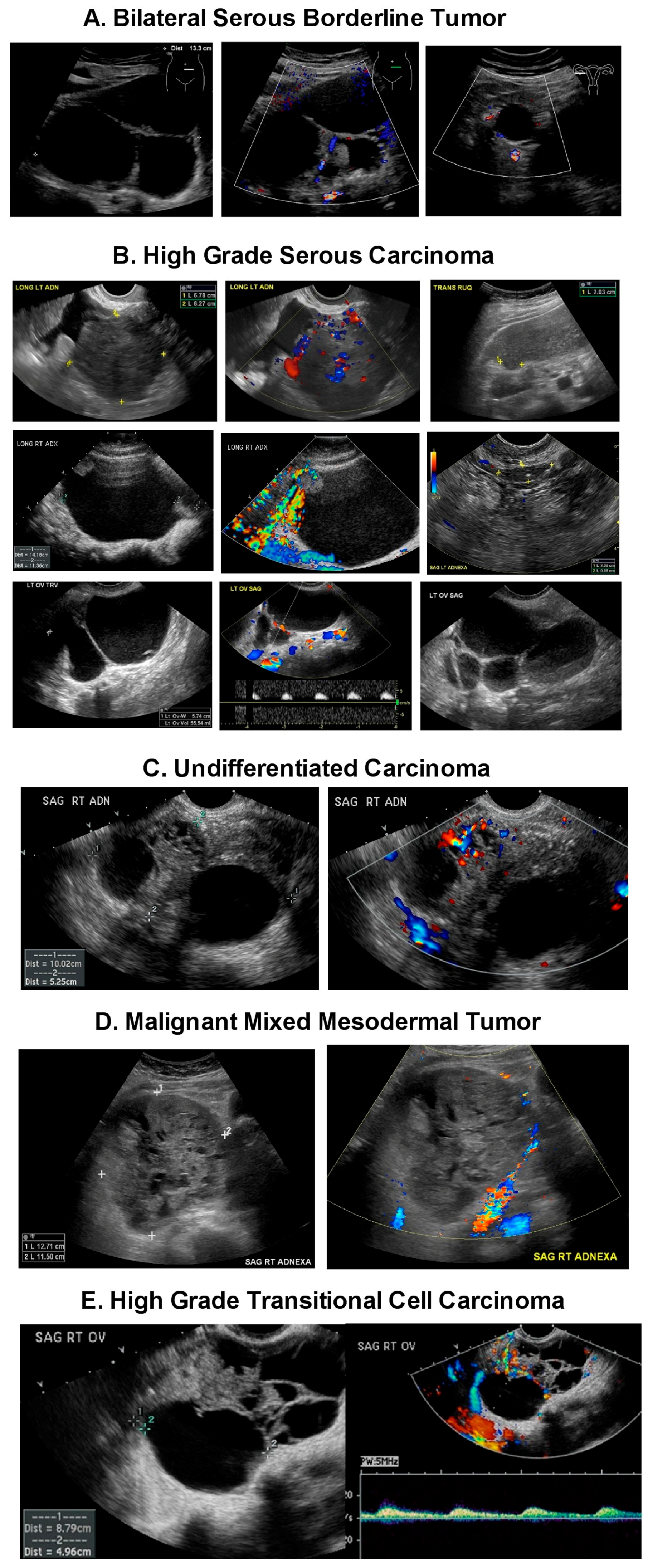
Diagnostics | Free Full-Text | Ultrasound Monitoring of Extant Adnexal Masses in the Era of Type 1 and Type 2 Ovarian Cancers: Lessons Learned From Ovarian Cancer Screening Trials | HTML
Ovarian cancer screening | CMAJ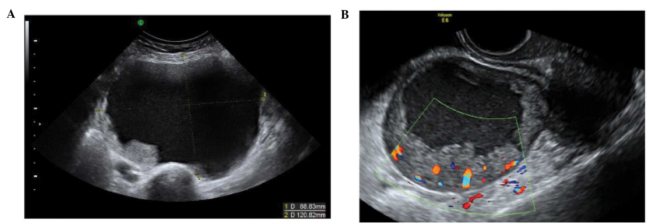
The characteristic ultrasound features of specific types of ovarian pathology (Review)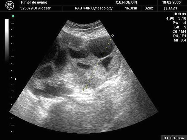
Malignant Ovarian Tumors (Serous/Mucinous/Endometrioid/Clear Cell Carcinoma): Clinical Setting and Ultrasound Appearance | SpringerLink
Simple vs. Complex Ovarian Cysts: The Link to Ovarian Cancer | Empowered Women's Health
Figure 1 from arly detection of ovarian cancer by tumor-epithelium targeted molecular ultrasound imaging in association with corresponding serum markers | Semantic Scholar
Watchful waiting' with routine ultrasound safer than removing benign ovarian cysts, study suggests - USF Health NewsUSF Health News
The enhancement in ultrasound signal intensity from spontaneous ovarian... | Download Scientific Diagram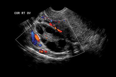
GPs 'refused access' to ovarian cancer scans | GPonline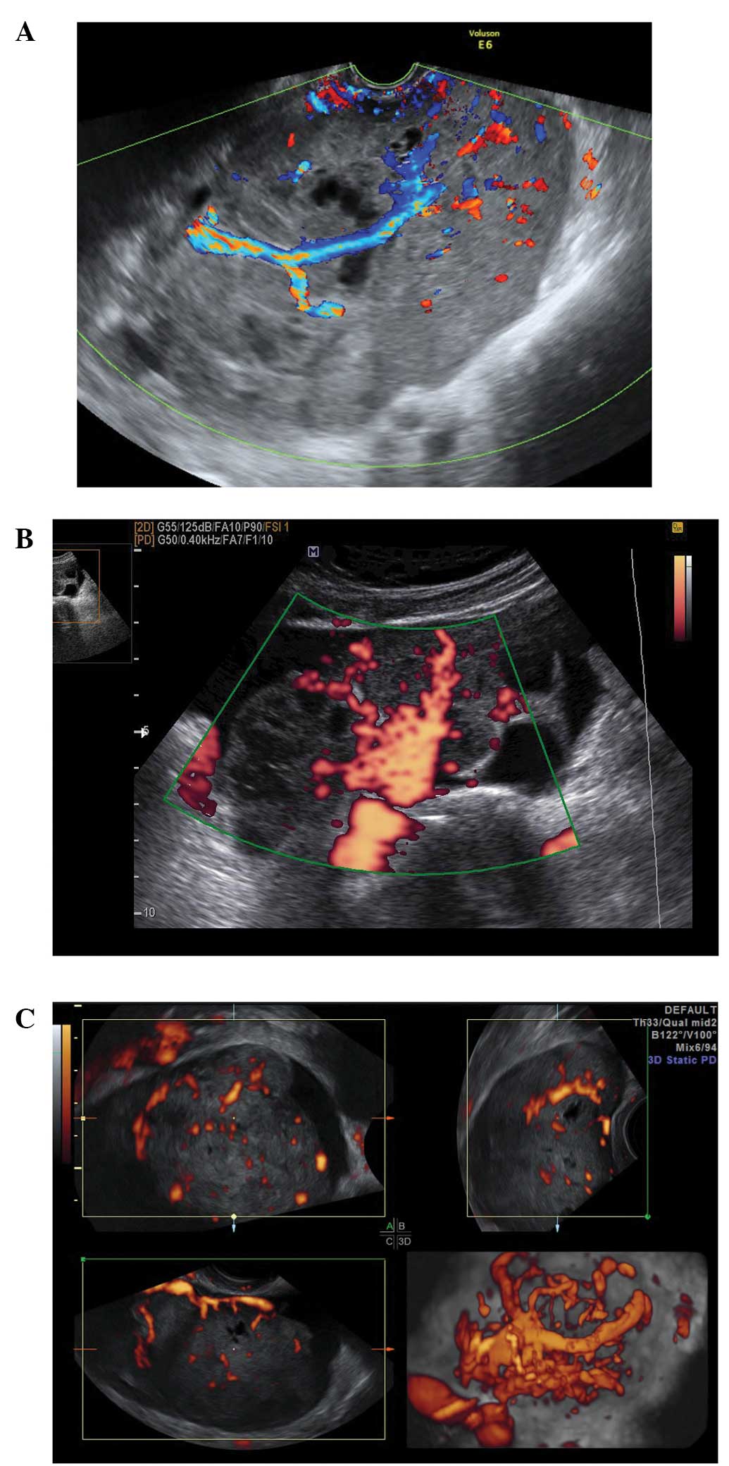
The characteristic ultrasound features of specific types of ovarian pathology (Review)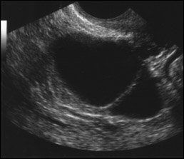
Some ovarian tumors can be safely followed on ultrasound
Pin on adnexa adnexa
Detection of spontaneous ovarian tumors at early stage with > v A 3... | Download Scientific Diagram
CEUS Identifies Ovarian Tumor – International Contrast Ultrasound Society
Ultrasound Video showing Metastatic ovarian cancer. - YouTube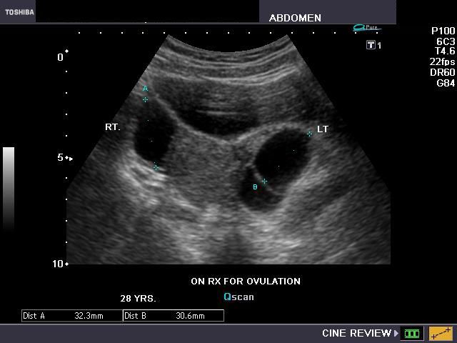
A Gallery of High-Resolution, Ultrasound, Color Doppler & 3D Images - ovaries
Ovarian cancer screening: shop for the story emphasis of your choice |
Simple vs. Complex Ovarian Cysts: The Link to Ovarian Cancer | Empowered Women's Health
A Better Way to Assess Ovarian Cancer Risk – NCAL Research Spotlight
Ultrasounds Improve Diagnosis for Ovarian Cancer | Clinical And Molecular Dx
IOTA Simple Rules and SRrisk calculator to diagnose ovarian cancer | Iota Group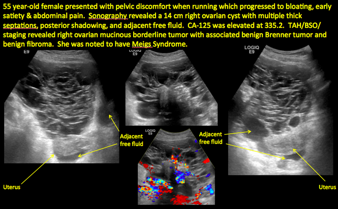
Imaging the suspected ovarian malignancy: 14 cases | MDedge ObGyn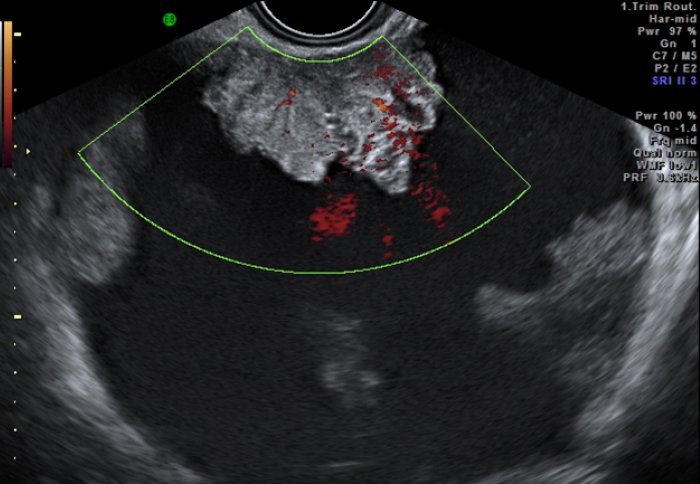
New test can help doctors choose best treatment for ovarian cancer | Imperial News | Imperial College London
Ultrasound Video showing an Ovarian tumor. - YouTube
Ovarian Tumors What is the clinical setting when you will consider an ovarian mass? You would consider an ovarian mass, in any woman who comes in complaining of pressure symptoms. These symptoms include urinary frequency, pelvic discomfort, and ...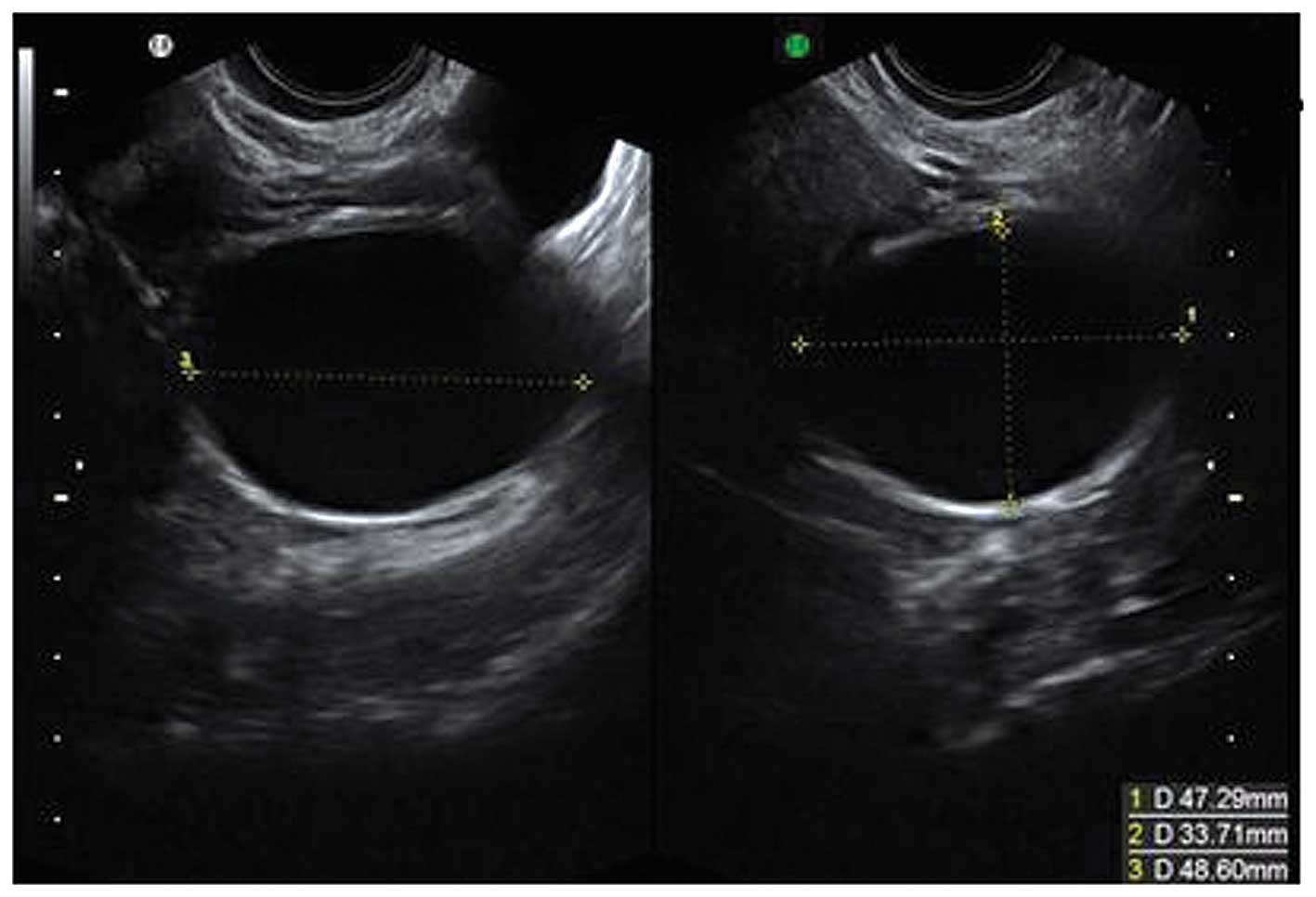
The characteristic ultrasound features of specific types of ovarian pathology (Review)
Diagnostic Ultrasound in the Assessment of the Adnexal Mass | GLOWM
Ovarian Cancer Pictures: Cysts, Symptoms, Tests, Stages, and Treatments
A Better Way to Assess Ovarian Cancer Risk – NCAL Research Spotlight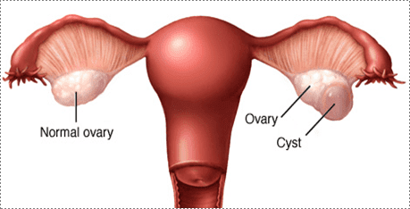
Ovarian Cysts and Ovarian Cancer: Alliance Obstetrics & Gynecology: Obstetricians
Watchful waiting' with routine ultrasound safer than removing benign ovarian cysts, study suggests - USF Health NewsUSF Health News
Immune Cells in the Normal Ovary and Spontaneous Ovarian Tumors in the Laying Hen (Gallus domesticus) Model of Human Ovarian Cancer


Posting Komentar untuk "what does ovarian cancer look like on an ultrasound"